
Sinus lift surgery
During the maxillary sinus lift surgery, sinus mucosa is lifted, then protected using a special membrane, and the sinus floor is filled using bone substitute.
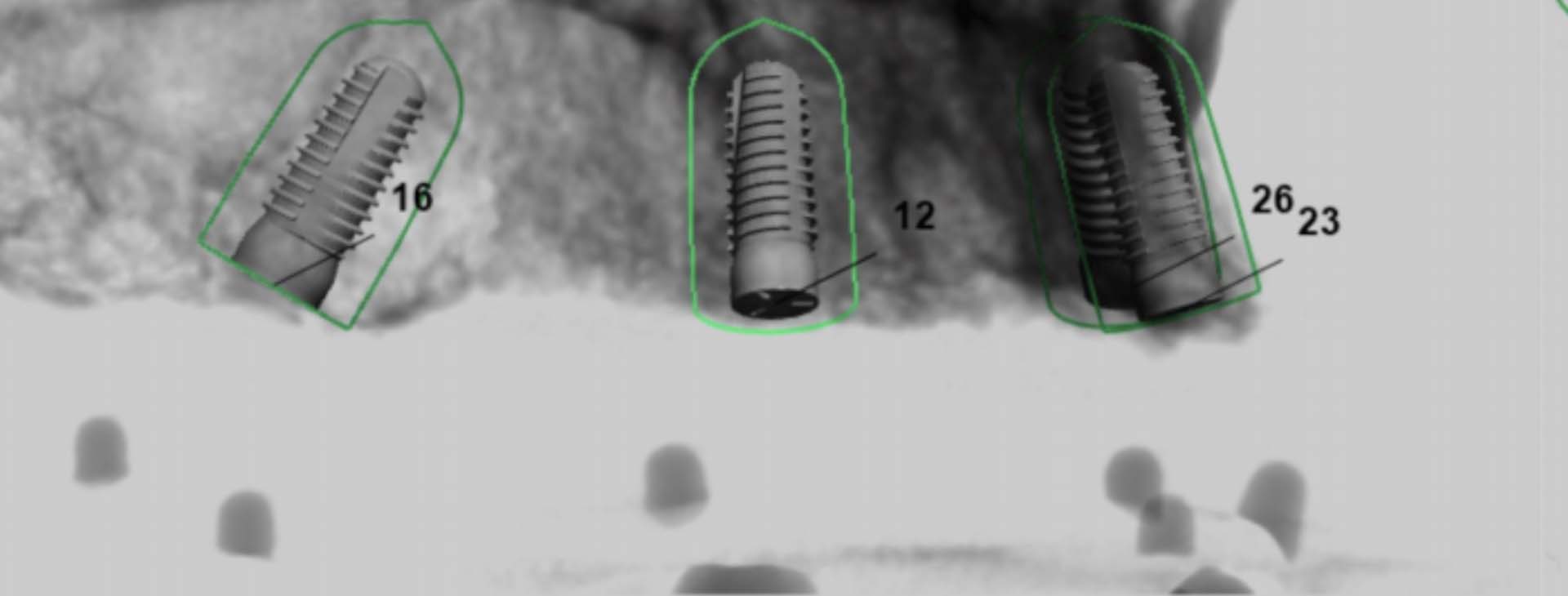
During the maxillary sinus lift surgery, sinus mucosa is lifted, then protected using a special membrane, and the sinus floor is filled using bone substitute. This way, the alveolar ridge height is increased to allow for implant placement to restore lost upper molars and premolars. Usually implants may be placed only 4 to 9 months after sinus lift surgery. This time is necessary for the placed bone substitute to heal, i.e., for blood vessels to grow into it and change it to regular bone. In some cases implants may be placed during the same visit.
During the consultation, your alveolar ridge height and width will be evaluated, you will be informed about the sinus lift surgery (open or closed technique), anesthesia (local, intravenous, general), preparation for the procedure, its complications and risks.
Preparation for sinus lift:
A panoramic X-ray and/or volumetric computed tomography is done before the procedure to evaluate the alveolar ridge form and anatomical structures. Important: it is advised to avoid flights and diving at least 7 days after the procedure.
No specific preparation is necessary for sinus lift under local anesthesia. Have a meal before the procedure, since it is not recommended to eat 2 to 3 hours after the surgery. If the surgery is planned under intravenous/general anesthesia, the following tests are recommended: Full blood test, blood glucose test, blood clotting tests (APTT, PT, INR), urine test and electrocardiogram (ECG).
Post-operative care:
1. It is normal to expect swelling, reddening, tenderness of the surgical site. Usually the swelling increases 2 to 4 days after the procedure.
2. There may also be pain upon touching the area. It usually starts to decrease after 3 to 4 days.
3. Bruising visible on the face. Usually most visible 2 to 4 days post procedure. Does not require any additional treatment. During resorption, the color of the bruise changes (from reddish blue to greenish yellow). The adjacent areas may also develop bruising. The size of the bruising depends on the scope and type of surgery and the body reaction to the procedure.


Other recommended treatments
The ProDentum clinic has a wide range of services and provides patients with the highest level of professionalism.